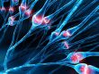
A new study published in Science has found that neurons in the brain do not follow a single strategy when learning. Instead, different parts of the same neuron—its upper and lower branches—adjust their connections using distinct rules. This finding challenges the traditional idea that each neuron has one learning rule, and it suggests that neurons may have more flexibility in how they process and store information.
Learning often involves modifying the strength of connections between neurons, a process known as synaptic plasticity. But scientists still don’t fully understand how the brain selects which connections to strengthen or weaken. This question, sometimes called the “credit assignment problem,” is important because only the right changes at the right time will lead to effective learning.
“The ability of our brains to adapt and learn from experience is fundamental for us to change our behavior in order to survive and succeed in the ever changing environments we live in. At the same time, some experiences can cause our brains to change in negative ways that then lead to behavioral deficits, such as what happens during addiction, depression, and PTSD. So understanding how our brains are able to adapt is not only critical to understand learning in general, but also has clinical implications,” explained study author William (Jake) Wright, a postdoctoral fellow in Takaki Komiyama’s lab at the University of California San Diego.
“Changes in the synaptic connections between neurons, which is how neurons communicate with each other, is thought to underlie learning by essentially changing how information flows through the brain. For this process of ‘synaptic plasticity’ to actually lead to learning, the right synaptic connections need to undergo the right types of changes at the right time. However it remains unclear how the brain selects which synapses to modify during learning, and this was the goal of our research.”
To investigate how neurons adjust their connections during learning, the researchers trained mice to perform a motor task. In the task, mice learned to press a lever in response to a tone in order to receive a water reward. As the mice practiced over the course of two weeks, they became faster and more consistent, showing that they had learned the task. Previous research had shown that this kind of motor learning leads to changes in the brain’s motor cortex, particularly in a type of neuron called a layer 2/3 pyramidal neuron.
The researchers focused on these neurons and used high-resolution imaging to watch changes in individual synapses—the small structures where one neuron receives input from another. Specifically, they used two advanced sensors: one to detect when glutamate, a chemical signal, was released onto a synapse, and another to measure calcium activity in the receiving neuron. These indicators allowed them to monitor both input to individual synapses and the overall output activity of the neuron.
They imaged two major parts of each neuron: apical dendrites, which extend toward the surface of the brain, and basal dendrites, which branch out closer to the cell body. These two regions have different structures and properties, raising the possibility that they might also follow different learning rules.
The results confirmed this hypothesis. The researchers found that synapses on apical dendrites tended to strengthen when they were active at the same time as nearby synapses. This means that local clusters of active inputs on these branches became stronger together, promoting what the researchers called functional clustering. In contrast, synapses on basal dendrites became stronger if they were active at the same time that the neuron fired an action potential—an electrical signal that represents output. This matches the classic view of learning in the brain, known as Hebbian learning, where connections strengthen when input and output are paired.
To test the importance of neuron output in this process, the researchers suppressed the ability of some neurons to fire action potentials. This manipulation had a strong effect on the basal dendrites: synapses in these branches no longer strengthened in response to training. However, the apical dendrites continued to show signs of learning through clustering, even when output activity was blocked. This showed that the two compartments not only followed different rules, but that only one of them—the basal dendrites—depended on output signals from the neuron to guide learning.
The study also revealed when these changes happened. Most of the strengthening of connections occurred in the early days of training, suggesting that this is a sensitive window during which the brain is most responsive to learning. The strengthening of synapses also tended to happen in neurons that later became more involved in movement control, linking the observed changes to behavioral outcomes.
By measuring how far apart the changing synapses were from one another, the researchers confirmed that apical dendrites tended to strengthen groups of nearby synapses, while basal dendrites showed no such clustering. This suggests that apical dendrites may be organizing information in a spatially grouped way, while basal dendrites may be more focused on aligning inputs with the overall activity of the neuron.
One of the key findings was that neurons can follow more than one learning rule at the same time, but apply those rules in specific parts of their structure. The apical dendrites appeared to operate based on local coactivity—when multiple nearby inputs are active together—while the basal dendrites responded to the coincidence of input and the neuron’s own output. These differences may arise from structural and electrical distinctions between the compartments. For example, signals from the rest of the brain may spread more easily into basal dendrites, while apical dendrites may be more electrically isolated, encouraging localized processing.
The study also raised questions about how these differences support learning. The researchers suggested that apical dendrites may group related inputs together, allowing the neuron to integrate complex information more efficiently. Meanwhile, the basal dendrites may help build associations between specific inputs and the neuron’s overall behavior. This dual strategy could allow neurons to perform more sophisticated computations and respond flexibly to different types of learning.
“We found that individual neurons utilize different synaptic plasticity rules across different sets of synapses depending on where those synapses are located on the neuron,” Wright told PsyPost. “This finding challenges the traditional view of synaptic plasticity, which often assumes plasticity rules are uniform and singular within a neuron.”
“Using multiple learning rules in tandem this way may allow neurons to perform distinct computations in parallel and grant them much greater computational capacity than typically thought. This finding may also have implications for the development of better and more efficient Artificial Intelligence, where most current neural networks use a single learning rule throughout.”
Although the findings provide new insight into how learning works in the brain, the authors noted some important limitations. The study was conducted in mice, and it focused on one type of neuron in one region of the brain. It is still unknown whether other neurons in different brain regions follow the same rules. More research is needed to determine how generalizable these findings are across species and brain systems.
The researchers plan to explore why neurons use multiple learning rules at once and how this flexibility benefits brain function. Understanding these processes may help scientists develop better treatments for neurological and psychiatric conditions where learning mechanisms go awry.
“One big remaining question is why do neurons utilized multiple learning rules at the same time,” Wright explained. “What functions and computations does this allow the neural circuits in our brains to perform?”
The study, “Distinct synaptic plasticity rules operate across dendritic compartments in vivo during learning,” was authored by William J. Wright, Nathan G. Hedrick, and Takaki Komiyama.